CRPS Images

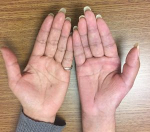
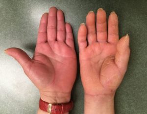
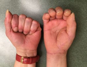
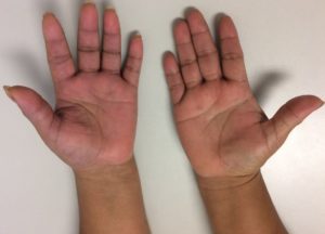
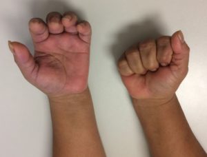
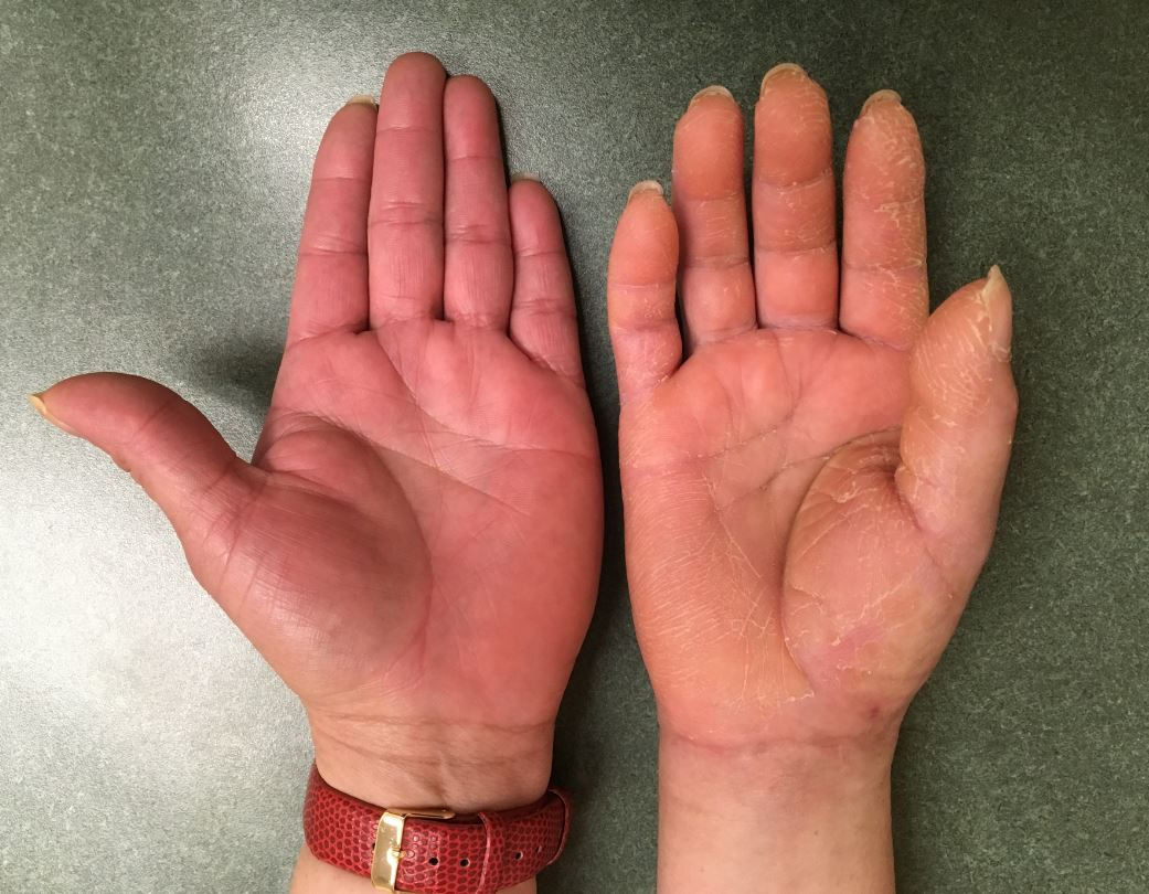
The following images represent classic signs and symptoms of Complex Regional Pain Syndrome, also known as CRPS.  Historically, this has also been referred to as sympathetic reflex dystrophy or causalgia.

I have found that the most sensitive and predictive finding is a dramatic loss in function (strength or motion) of a body part that is out of proportion to the injury.  Other common findings include:

  • changes in skin texture on the affected area; it may appear shiny and thin
  • abnormal sweating pattern in the affected area or surrounding areas
  • changes in nail and hair growth patterns
  • stiffness in affected joints
  • problems coordinating muscle movement, with decreased ability to move the affected body part
  • abnormal movement in the affected limb, most often fixed abnormal posture (called dystonia) but also tremors in or jerking of the limb.

Treatment always requires a prompt diagnosis and typically starts with therapy to improve motion and reduce discomfort.  However, some evidence suggests CRPS is propagated by a compressed nerve (ie: median nerve) and surgical intervention is sometimes recommend, such as a carpal tunnel release.

The following images show patients with an open palm, and attempted closed fist, compared to the normal side.

Mallet Finger CRPP

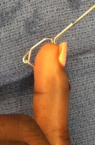
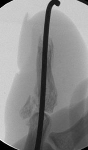
History: 31 year old female administrator injured her finger about 3 weeks prior to seeing me in the office.  She had a fracture-dislocation of the finger tip, also known as a mallet finger or mallet fracture with dislocation. I recommend surgical treatment.  To avoid making an incision, I recommended percutaneous pin treatment. 
Diagnosis: Right ring finger mallet fracture dislocation

Treatment: Percutaneous reduction with pinning.  Intra-operative technique shown here. 


Outcome: The small fragment pin was removed in the office after 6 weeks.  The longitudinal pin was removed after 10 weeks because she could not make an earlier appointment.  She had no pain, fully healed, and was using her finger normally.  By her last visit, some bridging bone could be seen across the fracture. 


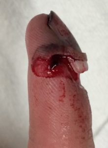
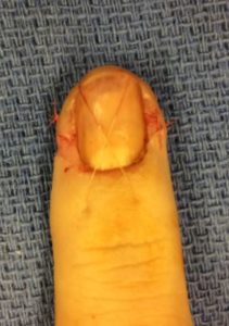
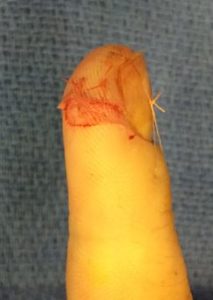
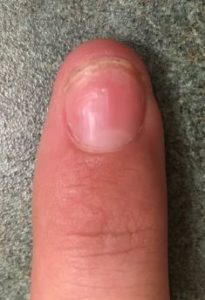
Nail Bed Avulsion

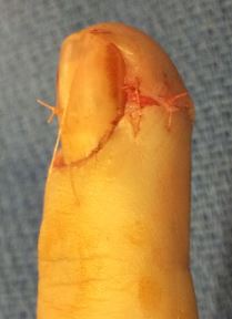
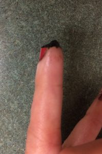
History: 18 year old male injured his index finger after dropping a 60-pound weight at the gym.  He presented to the emergency department for an open injury with an avulsed nail and nail bed laceration.  

Diagnosis: Right index finger open distal phalanx fracture with nail bed laceration and nail avulsion

Treatment: Repair of nail bed and replacement of nail plate

Outcome: He had a fast recovery.  The nail bed healed beautifully and the nail plate grew back without any abnormality.  He was back in the gym within a few weeks and by 3 months had a normal appearance of his finger.

Mucous Cyst Bilobed Flap

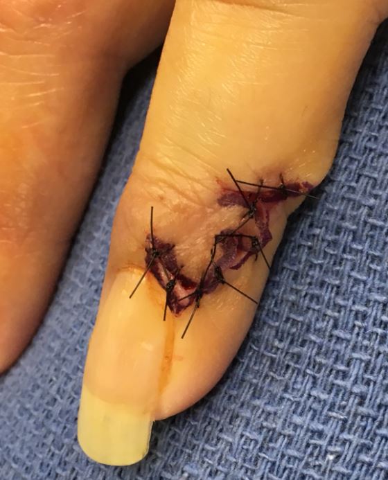
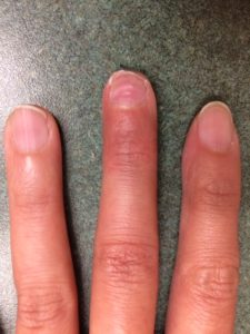
History: 44 year old female with a chronic mass at her fingernail.  The mass had been present for many months and would occasionally become infected and drain.  It was also causing a depression in her nail and quite uncomfortable and unsightly.  

Diagnosis: Left ring finger mucous cyst

Treatment: Mucous cyst excision with bilobed advancement flap

Outcome: She healed the incision beautifully and by 4 weeks the area was painless and improved. At her 3 month visit the nail plate and returned to normal appearance and she was very pleased. 

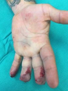
Finger Crush – Franko

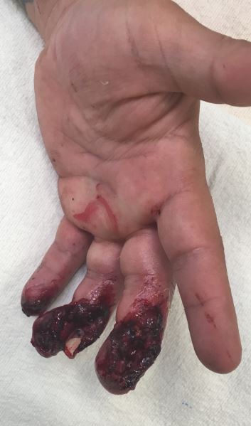
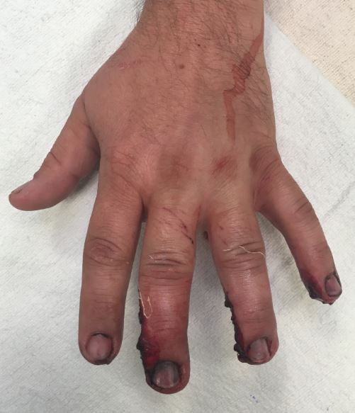
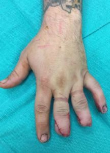
History: 37 year old gentleman was in a motorcycle accident where he sustained a severe trauma and crush to his middle and ring fingers.  I met him in the trauma bay where he had significant crushed finger tips and open wounds.  Based on the amount of injury, I recommended revising the amputated digit tips to regain function.

Diagnosis: Left 3rd and 4th digits open distal tuft fractures


Treatment: Revision amputation of the 3rd and 4th digits

Outcome: He had a rapid and smooth recovery.  He worked hard on therapy and regained function and strength within a few months.  Images below are from his 5 month appointment where he had returned to fully normal function. 

Fingertip V-Y

History: 39 year-old female who amputated the tip of her left middle finger while doing her hobby of leather crafting.  An emergency department cauterized the end of her finger to stop the bleeding and Dr. Franko saw her in the office for treatment.  She had loss of the soft tissue with exposed distal phalanx. 

Diagnosis: Left long finger tip amputation with exposed bone

Treatment: Operative debridement with V-to-Y advancement flap

Outcome: At 3 weeks after surgery the fingertip flap was healing and she was working on motion. Pain was minimal. 

By 3 months the fingertip looked natural and the nail was growing back normally.  She has full range of motion and minimal tenderness. 

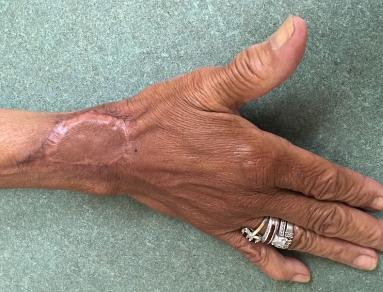
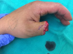
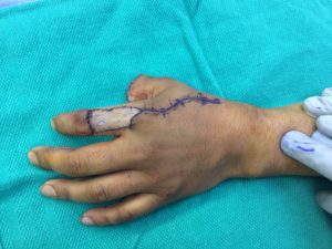
Thumb Amp FDMA Flap

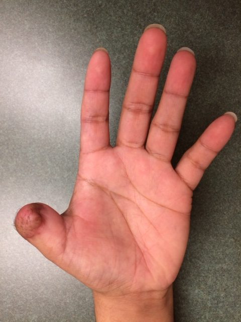
History: 25 year-old male injured his left thumb using a table saw resulting in an amputation of the thumb at the level of the interphalangeal joint. X-rays showed a partial thumb amputation.  He came to the emergency department with pain, disability, and bleeding. 

Diagnosis: Left thumb amputation at interphalangeal joint

Treatment: First dorsal metacarpal artery island flap

Outcome: He developed full healing and full use of his thumb within 2 months without difficulty.