CRPS Images

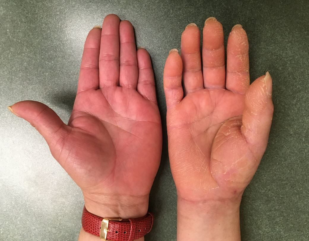
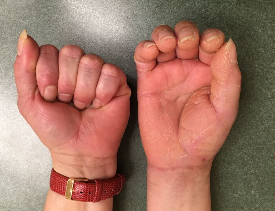
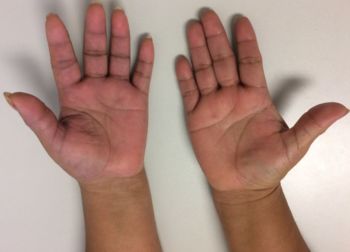
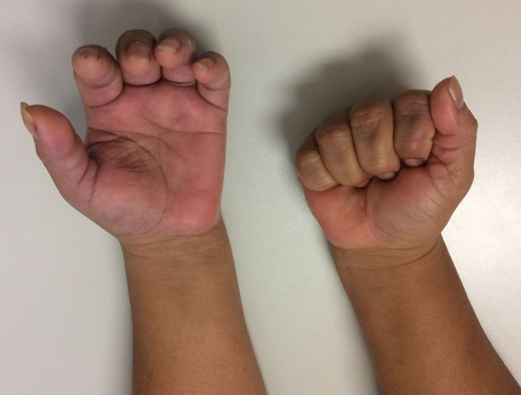
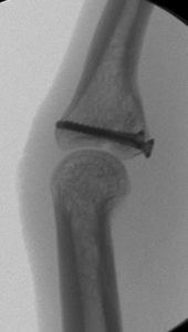
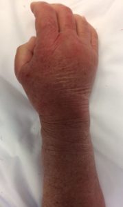
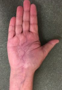
The following images represent classic signs and symptoms of Complex Regional Pain Syndrome, also known as CRPS.  Historically, this has also been referred to as sympathetic reflex dystrophy or causalgia.

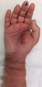
I have found that the most sensitive and predictive finding is a dramatic loss in function (strength or motion) of a body part that is out of proportion to the injury.  Other common findings include:

  • changes in skin texture on the affected area; it may appear shiny and thin
  • abnormal sweating pattern in the affected area or surrounding areas
  • changes in nail and hair growth patterns
  • stiffness in affected joints
  • problems coordinating muscle movement, with decreased ability to move the affected body part
  • abnormal movement in the affected limb, most often fixed abnormal posture (called dystonia) but also tremors in or jerking of the limb.

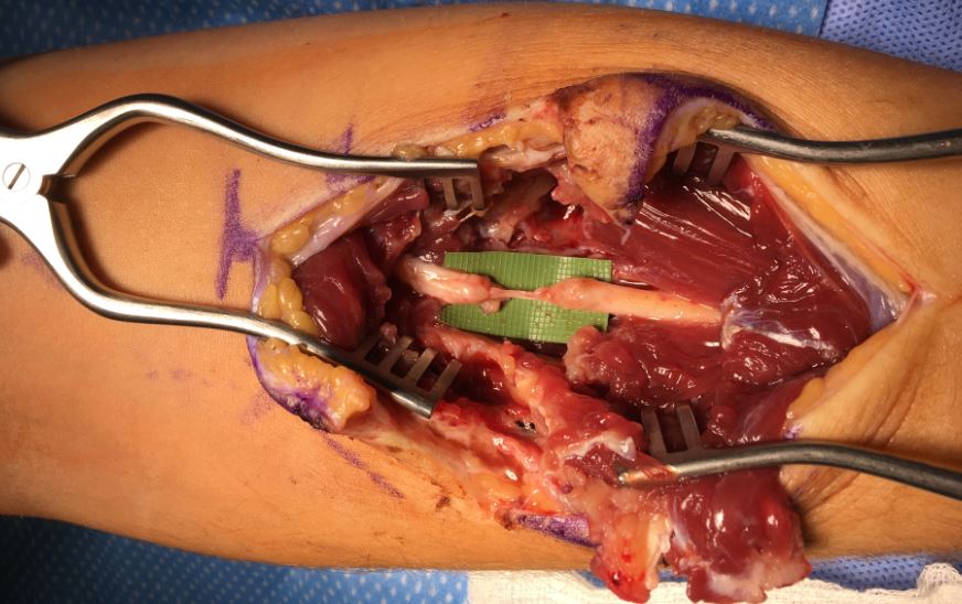
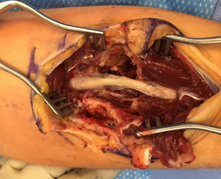
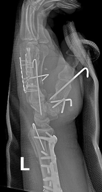
Treatment always requires a prompt diagnosis and typically starts with therapy to improve motion and reduce discomfort.  However, some evidence suggests CRPS is propagated by a compressed nerve (ie: median nerve) and surgical intervention is sometimes recommend, such as a carpal tunnel release.

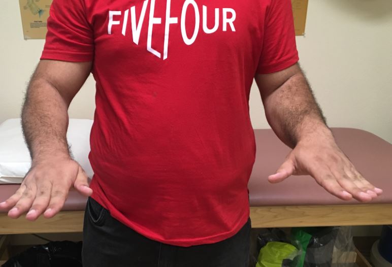
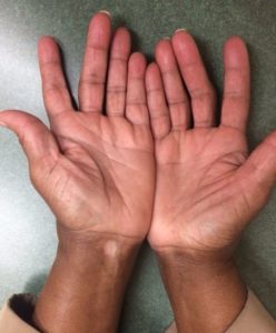
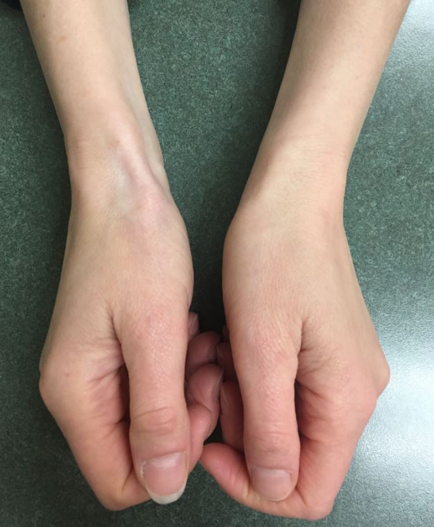
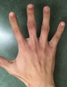
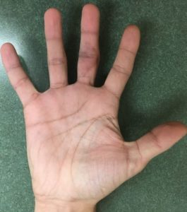
The following images show patients with an open palm, and attempted closed fist, compared to the normal side.

Median Nerve Allograft

History/Diagnosis: This patient is a 35 year old gentlement who sustained a glass puncture injury to his forearm about 3 weeks before presenting to my office for evaluation.  He initially did not think the injury was severe, but due to continued pain and numbness was concerned that it may be more involved.  His exam was consistent with numbness in the median nerve distribution and a laceration suggesting a median nerve injury. I recommended exploration and allograft repair. 

Treatment: Intra-operatively he was found to have a completed laceration of the median nerve.  I resected the nerve to healthy tissue and performed an allograft repair with suture and fibrin glue.


Outcome: He did very well during his recovery.  He recovered nearly 100% of his finger motion.  His sensation improved every visit and by 6 months he had sensation and tingling into the tips of his thumb, index, and middle fingers. 


Complex Motorcycle Accident

History/Diagnosis: This patient sustained a very severe motorcycle accident with open injuries to the forearm, wrist, hand, and fingers.  There were fractures of the ulna, radius, ulnar styloid, thumb dislocation, metacarpals, and CMC joints.  I took her to the operative room for extensive surgery to repair and reconstruct all of the injuries.  

Treatment: Complex repair of all injuries.

Outcome: She did exceptionally well, essentially regaining full function and strength.  She returned to riding motorcycles, returned to work, and was very pleased!


Radius Shaft Fracture ORIF

History/Diagnosis: This gentleman sustained an isolated radial shaft fracture. 

Treatment: Radial shaft ORIF

Outcome: He regained full motion and strength without any limitations


Bilateral Forearm ORIF

History: 22 year-old male injured in a motorcycle accident with bilateral forearm fractures of the radius and ulna.  

Diagnosis: Complex bilateral forearm fractures with comminution, open injuries, and need for repair


Treatment: Bilateral forearm ORIF


Outcome: Within 6 weeks he had near full motion. He had minimal pain and was quite pleased with the final result. 

Steroid Skin Blanching

The following images demonstrate the temporary depigmentation and atrophy effects of Kenalog injected in the hand and wrist. The follow-up image is 6 months later.


3 Phalanx IM screws

History: 39 year old gentleman came to the office after a steel beam fell on his hand, crushing his index, middle, and ring fingers.  He had fractures of the proximal phalanx to all three fingers and limited motion, swelling, and pain. 

Diagnosis: P1 fractures of index, middle, ring fingers


Treatment: Index and Middle fingers intramedullary screw, Ring finger pinning

Outcome: At 2.5 months he had nearly full motion and no pain.  The index and middle fingers were significantly better than the ring finger in regards to motion. 

Table Saw Skin Graft

History: 63 year old gentleman sustained a table saw injury to his right index finger.  This included a deep skin injury with full thickness loss but did not seem to involved the tendons, nerves, or vessels.  

Diagnosis: Right index full thickness loss

Treatment: Full thickness skin graft to digit

Outcome: Despite early loss of part of the graft, the entire wound quickly granulated in resulting in full coverage, full motion, and return to full activities. 

PIP Joint ORIF

History: A 19 year old gentleman injured his middle finger joint while punching a wall.  He presented to my office with an inability to flex and extend his middle finger and with pain and swelling. 

Diagnosis: Middle finger PIP joint intra-articular fracture with subluxation, moderately improved with traction

Treatment: Middle phalanx open reduction, internal fixation with screws

Outcome: He did exceptionally well.  At his first post-op visit he could nearly make a full fist and within 2 months he had nearly full motion without any pain. 

Deep Palm Infection

History:  71 year old female presented to the hospital about 2 weeks after a fall with increasing pain and swelling in her hand and palm. She had a history of psoriatic arthritis for which she took methotrexate and infliximab.  Her presentation was very concerning for a deep infection with a WBC of 41,000, lactate of 2.6, and a CT scan in the emergency department showing a deep abscess around the median nerve. 

Diagnosis: Left palm deep space infection in the setting of immunosuppresion

Treatment: Urgent I&D of the deep space through an extended carpal tunnel incision, before and after photos

Outcome: She made a rapid recovery with IV antibiotics. Ultimately she was able to regain nearly all of her function and return to her usual daily routine.