Lunate Facet Malunion with Osteotomy

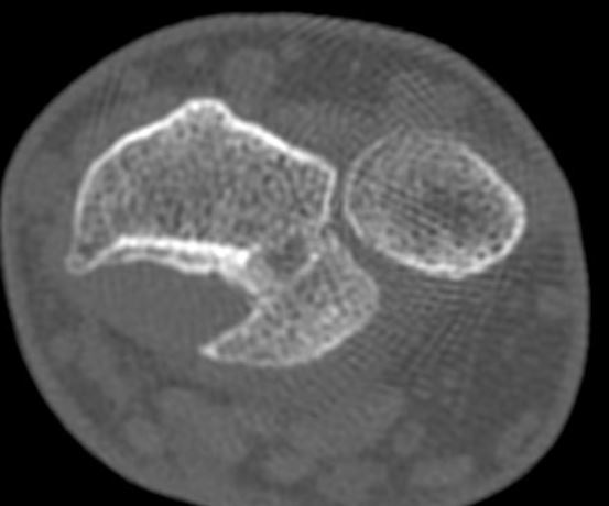
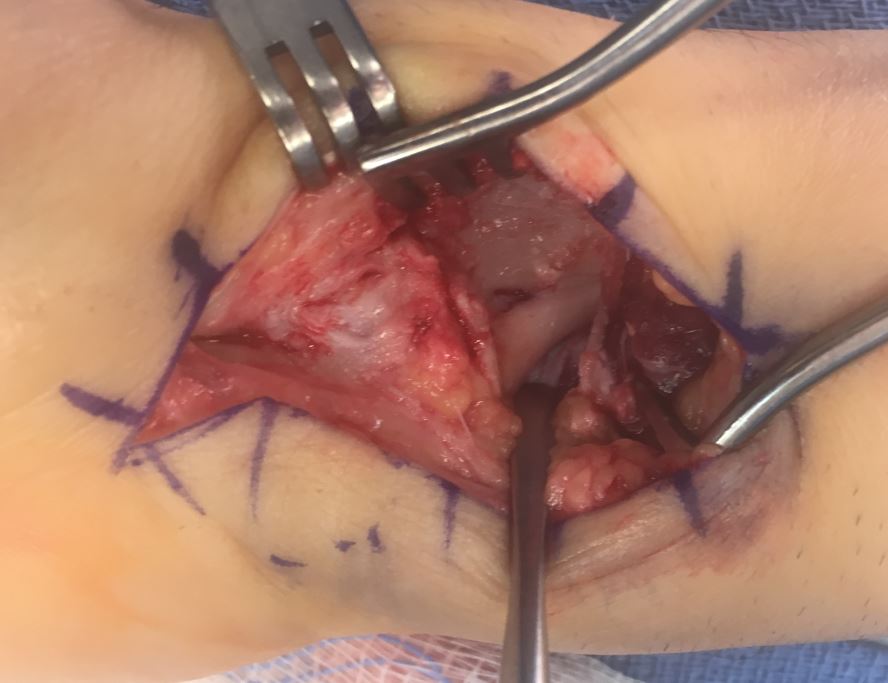
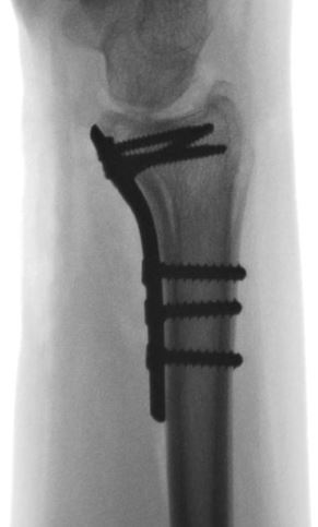
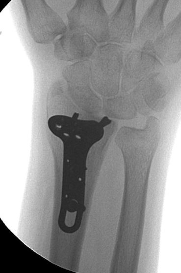
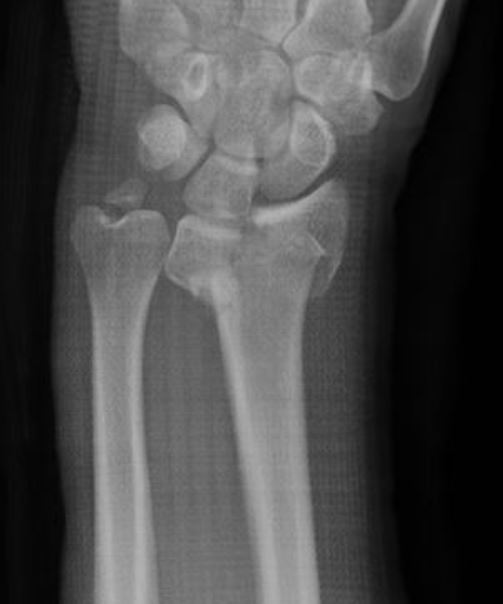
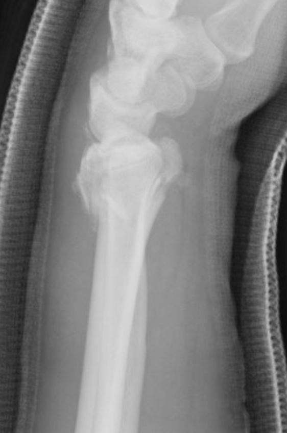
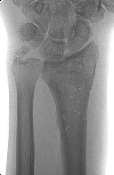
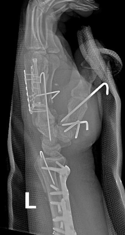
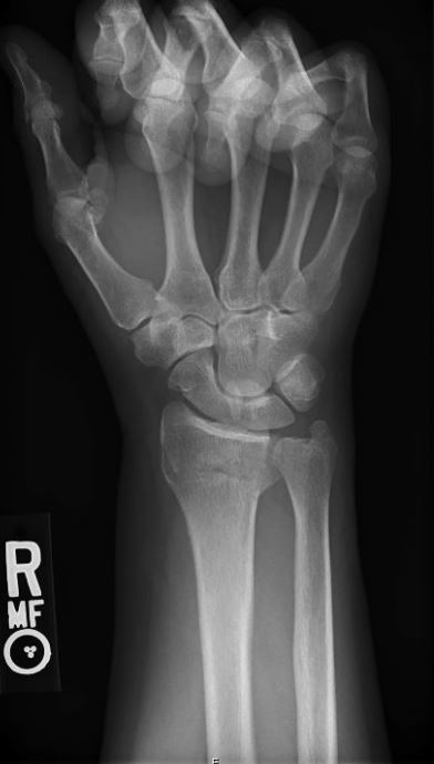
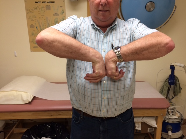
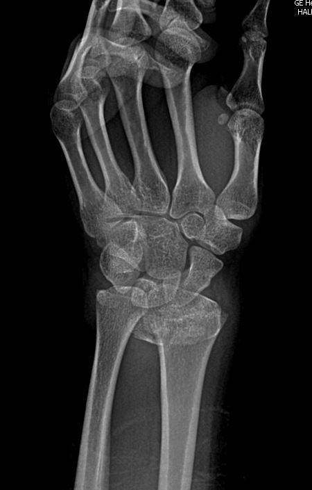
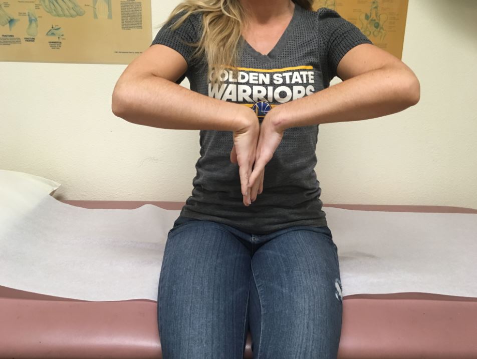
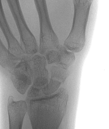
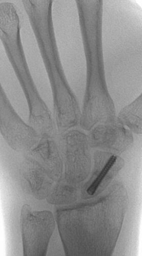
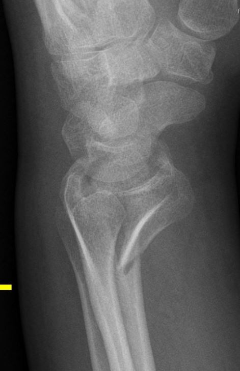
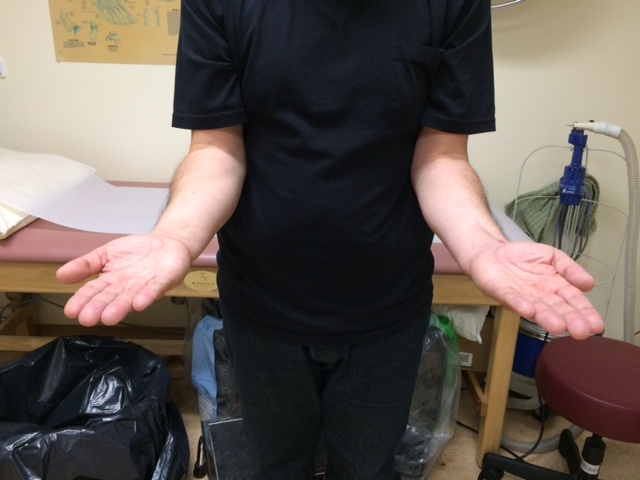
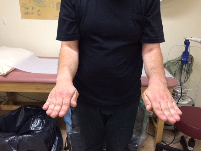
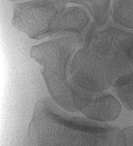
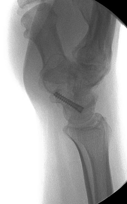
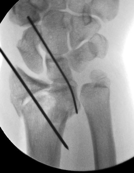
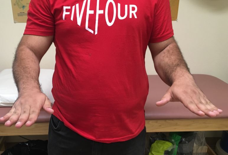
Case Presentation: A 36 year old gentleman sustained a wrist injury while running and falling but xrays at the time were reportedly negative for a fracture.  He was given a splint but had continued and persistent pain and swelling.  He came to my office 6 months later and xrays and a CT scan showed a dislocated wrist with a malunited lunate facet fragment.  Based on his young age and long term function, I recommended an osteotomy to correct the bone and restore the articular surface.  The fracture healed uneventfully and he worked aggressively with therapy to regain motion which plateaued at 1 year with wrist flex/ext of 25/55 degrees and supination/pronation of 50/80 degrees.


Distal Radius 3 plates

Case Presentation: 30 year old gentleman injured his distal radius (wrist fracture) while snowboarding.  He had a severe fracture with separation of the lunate and scaphoid facet fragments.  Intra-operative fixation required individual fragment specific plating with screws, wires, and plates.  The fracture healed in a reasonable position, and the plates were taken out once healed due to some mild irritation. Once recovered he had nearly full motion and was very pleased.


Complex Motorcycle Accident

History/Diagnosis: This patient sustained a very severe motorcycle accident with open injuries to the forearm, wrist, hand, and fingers.  There were fractures of the ulna, radius, ulnar styloid, thumb dislocation, metacarpals, and CMC joints.  I took her to the operative room for extensive surgery to repair and reconstruct all of the injuries.  

Treatment: Complex repair of all injuries.

Outcome: She did exceptionally well, essentially regaining full function and strength.  She returned to riding motorcycles, returned to work, and was very pleased!


Segmental Galeazzi

History: 24 year old gentleman presented to the hospital after crashing his motorcycle.  He had obvious deformities to his right forearm and wrist.  I evaluated him and recommended surgery for his complex radial shaft and DRUJ injuries. Intra-operatively he was found to have multiple shaft fractures, further complicating the surgery. 

Diagnosis: Right wrist and forearm Galeazzi fracture

Treatment: Intra-operative findings necessitated and combination of pins and plate extensions in addition to standard volar plating and screws to capture multiple fracture components.  The sequence of fixation is shown here. 

Outcome: He did very well in the early post-operative period, making improvements in his wrist motion and forearm supination.  By 1 month he had already regained 30% of wrist motion and 50% of forearm supination.  He had very little pain and was slowly returning to activity. 

Distal Radius Osteotomy

History: 55 year old female had sustained a left wrist fracture 3 months prior to coming to my office.  A different surgeon treated her with a cast for 6 weeks followed by brace.  For the prior 2 months however she had progressive pain, swelling, and numbness/tingling in her wrist and fingers suggestive of carpal tunnel syndrome.  X-rays showed the fracture did not heal properly and she was very bothered by it.  She requested the distal radius be fixed. 

Diagnosis: Left distal radius malunion with carpal tunnel syndrome

Treatment: Distal radius osteotomy through volar approach with iliac crest bone graft and carpal tunnel release

Outcome: Over the first 2 weeks after surgery she had immediate improvement in her carpal tunnel symptoms.  As the pain improved, she regained full motion and strength in her wrist.  All of her pain was alleviated and she was very happy by 3 months after surgery. 


Distal Radius ORIF

History: 62 year-old male fell in his backyard injuring his right wrist.  X-rays showed a wrist fracture with displacement and angulation resulting in severe pain, swelling, and deformity.

Diagnosis: Right distal radius fracture

Treatment: Open reduction and internal fixation with plates and screws

Outcome: At 6 weeks after surgery he had regained nearly all motion, was still working on increasing his strength, and had no pain.  The scar was hardly noticeable. X-rays showed the bones were in perfect alignment.

No pain and nearly normal motion at 6 weeks after surgery! 

Wrist Fracture (severe)

History: 24 year-old female Warriors dancer fell at work and injured her left wrist.  She presented to the hospital and was diagnosed with a severe distal radius fracture (wrist fracture).  After undergoing a closed reduction (re-alignment of the bones) in the emergency room, Dr. Franko recommended surgery to provide strength and stability to the bones and allow them to heal properly. 

Diagnosis: Left intra-articular distal radius fracture with ulna styloid fracture


Treatment: Left distal radius open reduction internal fixation with tension suture for ulna styloid

Outcome: At her first post-operative visit she transitioned immediately to a removable splint and began working with hand therapy on motion and strength.  Her scars healed quickly and she regained motion rapidly. 

At only 10 weeks from surgery she is fully healed with nearly normal motion and strength.  She will return to dancing for the Warriors just in time for playoffs!


Her incisions are healed beautifully!

Scaphoid ORIF

History: 23 year-old male who came to my office with pain in his wrist about 3 months after a fall. At the time he thought he only sprained his wrist. X-rays in the office showed a scaphoid non-union (not healed) causing his pain. Due to concern about long-term development of arthritis and pain, he elected to have surgery for treatment.

Diagnosis: Left scaphoid non-union, 3 months

Treatment: Scaphoid open reduction internal fixation with bone graft for non-union

Outcome: By 4 months he was fully healed with minimal pain and near full motion. He was very happy with the result.

DRFx Wire under Plate

History: 46 year-old male who was injured while skateboarding on Christmas Eve. He presented to the hospital with severe pain and deformity in his left wrist. X-rays showed a severe wrist fracture with intra-articular impaction, displacement and angulation resulting in severe pain, swelling, and deformity as well as a displaced ulnar styloid base fracture.

Diagnosis: Left intra-articular distal radius fracture and ulna styloid fracture

Treatment: Open reduction and internal fixation with a combination of plates, screws, and wires.  A photo from the operating room (below) shows the use of pins to hold two of the fracture fragments supported beneath the plate. 

Outcome: At 2 months he was doing very well with a healed fracture and working on improving his motion.  A view of the Right wrist is shown as a comparison for alignment of the joint surface.

He returned at 4 months for a final check and demonstrated full motion, no pain, and no limitation to any activities. He was very happy with his result. 

Scaphoid Non-Displaced

History: 52 year-old male who fell directly onto his right hand with pain and swelling. An initial X-ray was negative for fracture in the Emergency Department. He was evaluated by Dr. Franko who recognized a subtle scaphoid fracture and recommended either non-operative (cast) or operative (surgery) treatment. Because the patient desired a faster return to work, activities, and strengthening while avoiding a long-term cast treatment, he elected for surgery.

Diagnosis: Right scaphoid waist fracture


Treatment: Right scaphoid mini-open internal fixation

Outcome: By 6 weeks the fracture was healing and he was aggressively working on motion with therapy. He had minimal pain and had returned to full work. At 3 months a CT scan showed the fracture was fully healed and by 5 months he had fully regained motion without any pain or discomfort.