Metacarpal IM Screw

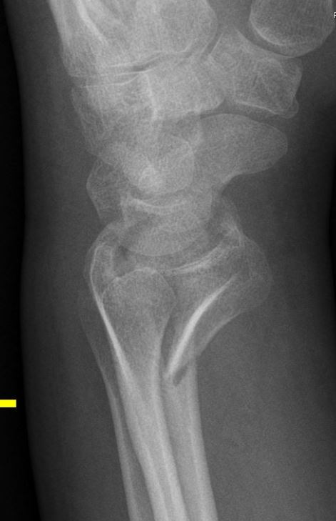
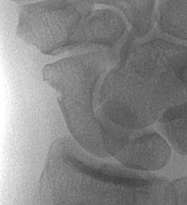
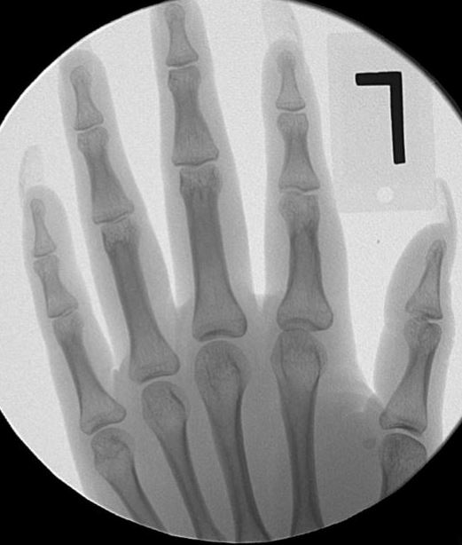
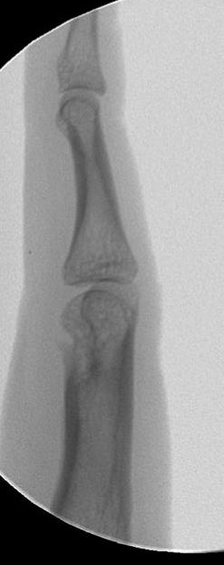
History: 30 year-old right-hand dominant male Sheriff impacted his left hand on concrete while wrestling a suspect.  He had immediate pain and swelling over the base of the index finger. X-rays showed a metacarpal fracture with displacement and angulation resulting in severe pain, swelling, and deformity.

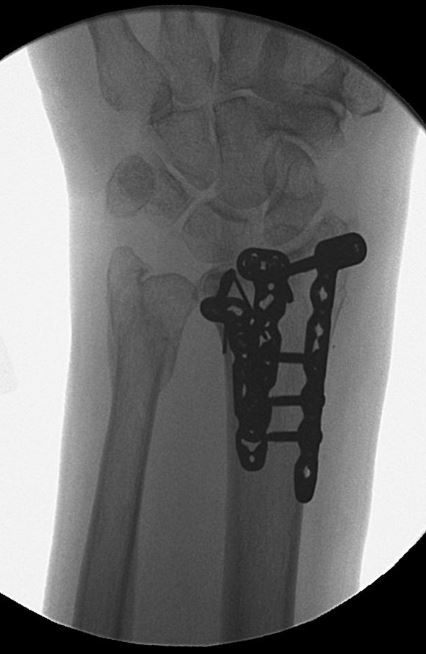
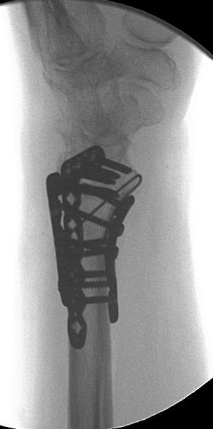
Diagnosis: Left 2nd metacarpal fracture, displaced

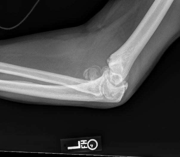
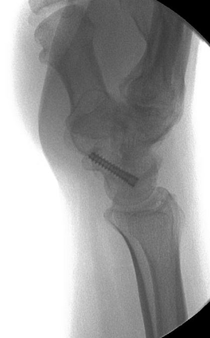
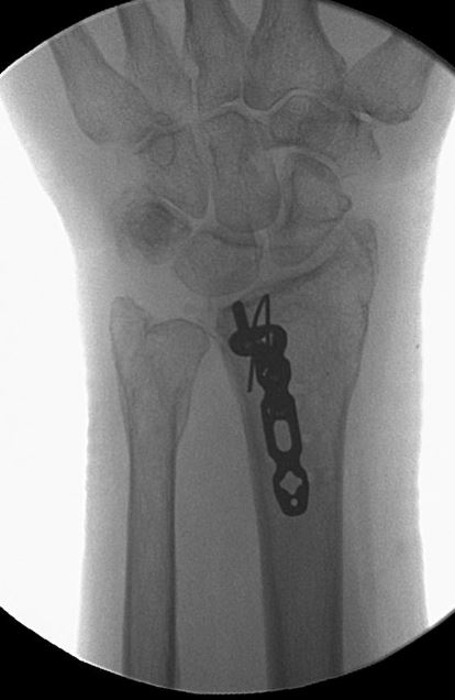
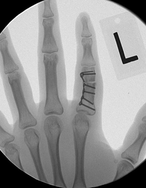
Treatment: Percutaneous reduction and internal fixation with an intramedullary screw

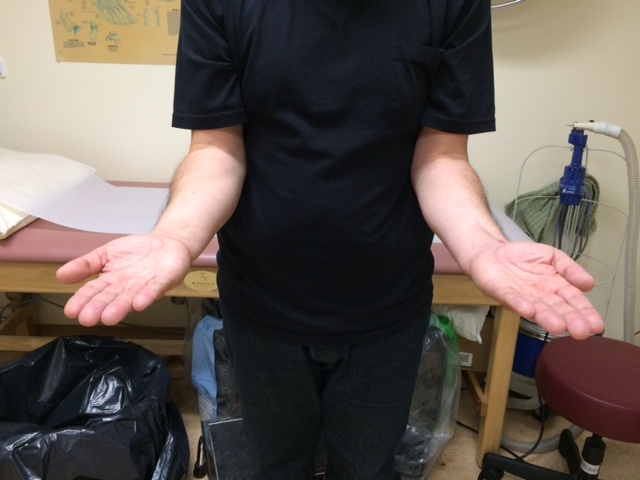
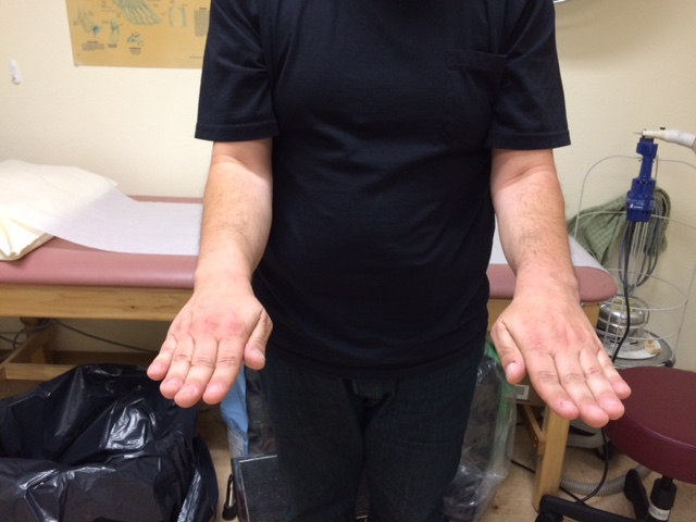
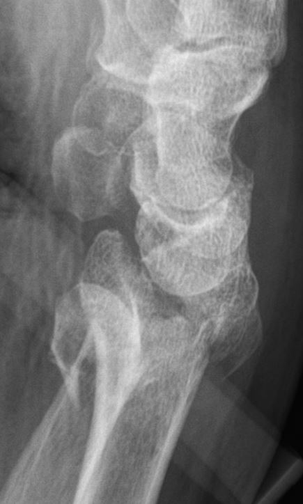
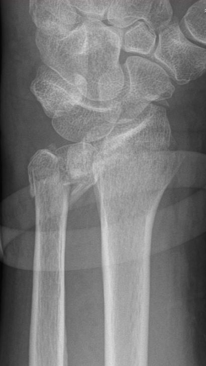
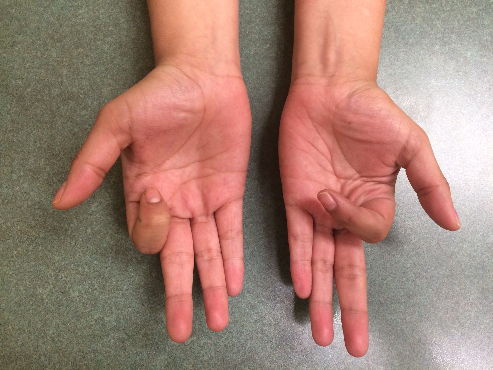
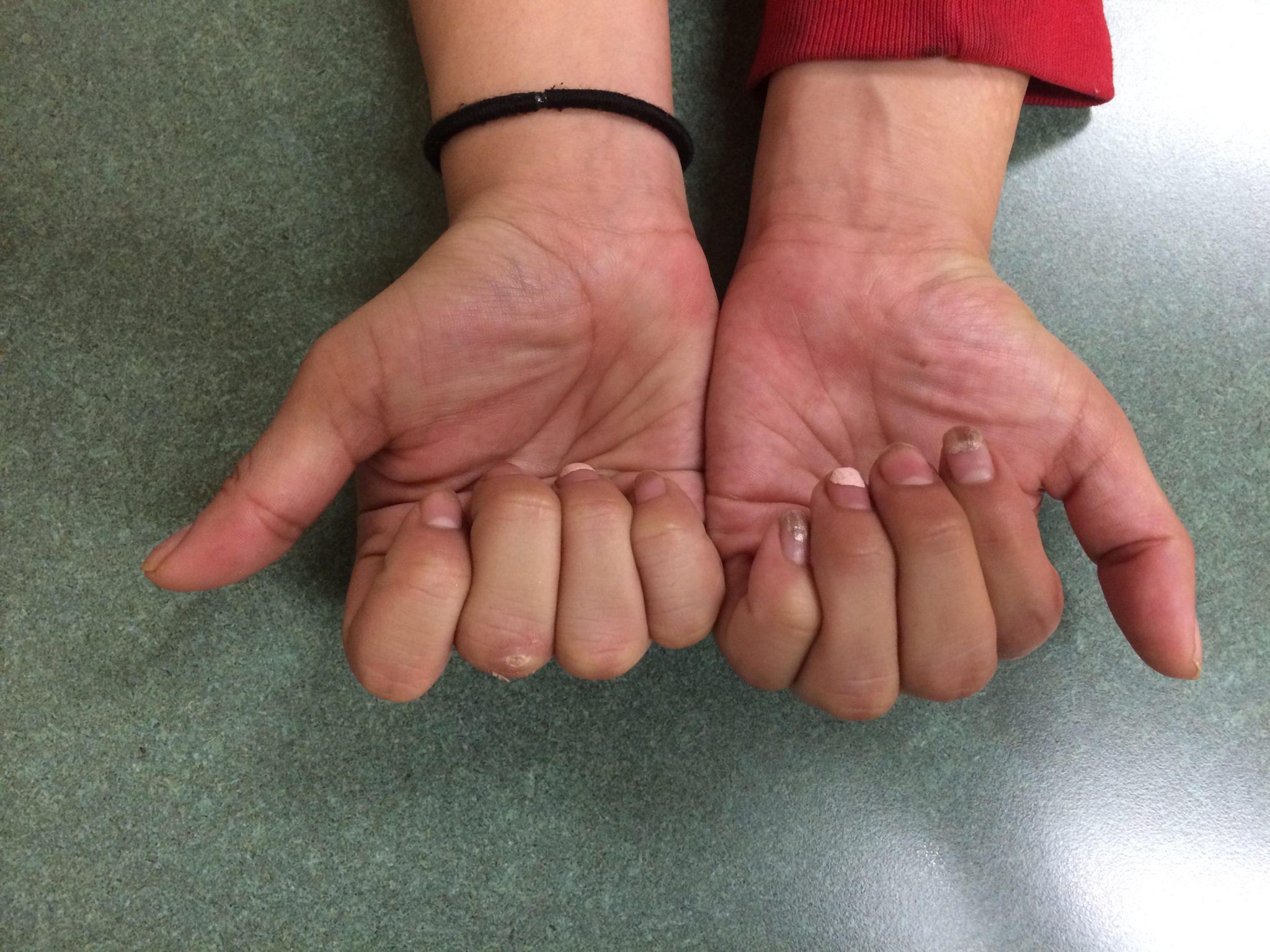
Outcome: He had immediate return of motion.  After 2 months of working with therapy, he had full healing, no pain, complete return of motion, and returned to work as a sheriff.  The scar was not noticeable. X-rays showed the bone was in perfect alignment.

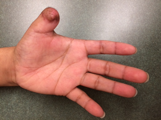
No pain and nearly normal motion at 2 months after surgery! 

DRFx Wire under Plate

History: 46 year-old male who was injured while skateboarding on Christmas Eve. He presented to the hospital with severe pain and deformity in his left wrist. X-rays showed a severe wrist fracture with intra-articular impaction, displacement and angulation resulting in severe pain, swelling, and deformity as well as a displaced ulnar styloid base fracture.

Diagnosis: Left intra-articular distal radius fracture and ulna styloid fracture

Treatment: Open reduction and internal fixation with a combination of plates, screws, and wires.  A photo from the operating room (below) shows the use of pins to hold two of the fracture fragments supported beneath the plate. 

Outcome: At 2 months he was doing very well with a healed fracture and working on improving his motion.  A view of the Right wrist is shown as a comparison for alignment of the joint surface.

He returned at 4 months for a final check and demonstrated full motion, no pain, and no limitation to any activities. He was very happy with his result. 

Distal Humerus

History: 60 year-old male who injured his left elbow while riding his bicycle.  He went to the hospital where he was diagnosed with a severe fracture of the elbow that included the joint surface.  He he also had some arthritis in the elbow prior to his fall but it was not very bothersome to him. 

Diagnosis: Left complex distal humerus fracture 

Treatment: Left elbow open reduction internal fixation through an olecranon osteotomy

Outcome: His recovery was very smooth. Within a few weeks he was already using his arms for crutches and working on his range of motion. By 4 months after surgery he had near full motion with no pain. After 6 months he was fully healed. The incision was nearly invisible.

Scaphoid Non-Displaced

History: 52 year-old male who fell directly onto his right hand with pain and swelling. An initial X-ray was negative for fracture in the Emergency Department. He was evaluated by Dr. Franko who recognized a subtle scaphoid fracture and recommended either non-operative (cast) or operative (surgery) treatment. Because the patient desired a faster return to work, activities, and strengthening while avoiding a long-term cast treatment, he elected for surgery.

Diagnosis: Right scaphoid waist fracture


Treatment: Right scaphoid mini-open internal fixation

Outcome: By 6 weeks the fracture was healing and he was aggressively working on motion with therapy. He had minimal pain and had returned to full work. At 3 months a CT scan showed the fracture was fully healed and by 5 months he had fully regained motion without any pain or discomfort.

DRFx Fragment Specific

History: 64 year-old female who was in a motor vehicle accident and admitted to the hospital for severe injuries including a fracture of the distal radius and distal ulna (wrist) on her left wrist. X-rays and CT scans showed a severe wrist fracture with intra-articular impaction, displacement and angulation resulting in severe pain, swelling, and deformity.

Diagnosis: Left distal radius fracture

Treatment: Open reduction and internal fixation with 3 plates, screws, and wires

Outcome: At 4 months she was doing well with a healed fracture but with persistent pain and irritation over the dorsal aspect of her wrist and limited motion.  After working hard with physical therapy, we agreed to removal the dorsal plates and screws to reduce discomfort.  

Just 1 month after removing the dorsal plates and screws she was already improved.  Scars were fully healed and her motion was almost back to normal.  Strength is still improving.  She is very happy with the result. 

P1 Derotation Osteotomy

History: 21 year-old female who fractured her left index finger in a car accident 2 years ago.  She was treated without surgery which resulted in the bone healing with a rotational deformity.  She notices that her index finger crosses over her other fingers when she makes a fist. 

Diagnosis: Left index proximal phalanx rotational malunion

Treatment: Left index finger proximal phalanx derotational osteotomy with locking blade plate

Outcome: After re-fracturing and fixing the bone in proper alignment, she no longer had “crossing over” of the index finger with flexion.  She worked hard with physical therapy and regained nearly all of her motion without any pain in the finger. 

Great motion and no pain about 4 months after surgery.  

Thumb Amp FDMA Flap

History: 25 year-old male injured his left thumb using a table saw resulting in an amputation of the thumb at the level of the interphalangeal joint. X-rays showed a partial thumb amputation.  He came to the emergency department with pain, disability, and bleeding. 

Diagnosis: Left thumb amputation at interphalangeal joint

Treatment: First dorsal metacarpal artery island flap

Outcome: He developed full healing and full use of his thumb within 2 months without difficulty.