PIP Joint ORIF

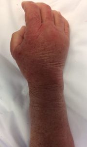
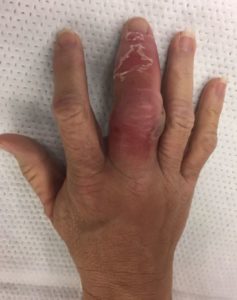
History: A 19 year old gentleman injured his middle finger joint while punching a wall.  He presented to my office with an inability to flex and extend his middle finger and with pain and swelling. 

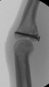
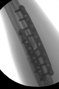
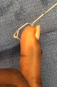
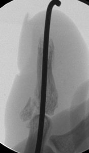
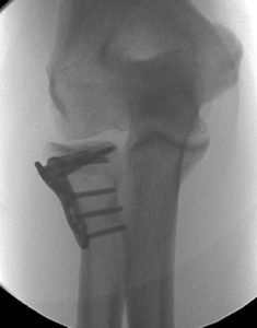
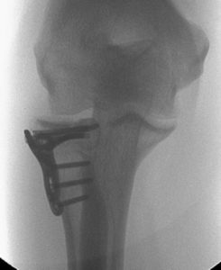
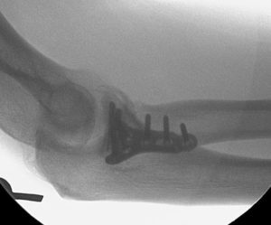
Diagnosis: Middle finger PIP joint intra-articular fracture with subluxation, moderately improved with traction

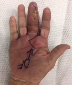
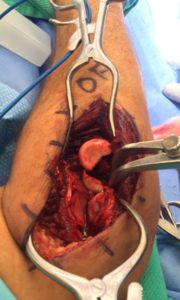
Treatment: Middle phalanx open reduction, internal fixation with screws

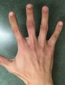
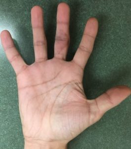
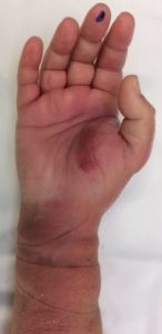
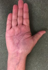
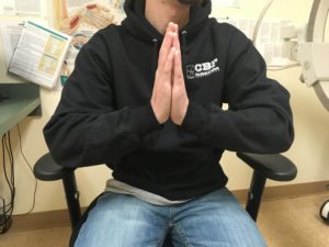
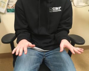
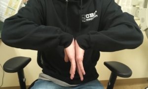
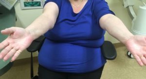
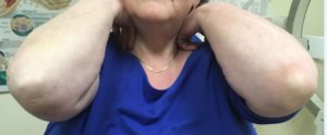
Outcome: He did exceptionally well.  At his first post-op visit he could nearly make a full fist and within 2 months he had nearly full motion without any pain. 

Deep Palm Infection

History:  71 year old female presented to the hospital about 2 weeks after a fall with increasing pain and swelling in her hand and palm. She had a history of psoriatic arthritis for which she took methotrexate and infliximab.  Her presentation was very concerning for a deep infection with a WBC of 41,000, lactate of 2.6, and a CT scan in the emergency department showing a deep abscess around the median nerve. 

Diagnosis: Left palm deep space infection in the setting of immunosuppresion

Treatment: Urgent I&D of the deep space through an extended carpal tunnel incision, before and after photos

Outcome: She made a rapid recovery with IV antibiotics. Ultimately she was able to regain nearly all of her function and return to her usual daily routine. 

Flexor Tenosynovitis

History: A 55 year old female with diabetes presented to my office with severe pain and swelling in her middle finger a few days after burning the tip of her finger.  This was initially treated with antibiotics for cellulitis however she rapidly progressed to flexor tenosynovitis based on the appearance and was taken to the operating room for urgent exploration, irrigation, and debridement of the tendon sheath. 

Diagnosis: Right middle finger flexor tenosynovitis

Treatment: Flexor sheath I&D followed by daily soaks

Outcome: Over weeks to months she healed the incision.  The flexor tendons required excision and she had limited flexion of the finger, but it remains infection-free.  She currently has very limited feeling in the finger. 

Segmental Galeazzi

History: 24 year old gentleman presented to the hospital after crashing his motorcycle.  He had obvious deformities to his right forearm and wrist.  I evaluated him and recommended surgery for his complex radial shaft and DRUJ injuries. Intra-operatively he was found to have multiple shaft fractures, further complicating the surgery. 

Diagnosis: Right wrist and forearm Galeazzi fracture

Treatment: Intra-operative findings necessitated and combination of pins and plate extensions in addition to standard volar plating and screws to capture multiple fracture components.  The sequence of fixation is shown here. 

Outcome: He did very well in the early post-operative period, making improvements in his wrist motion and forearm supination.  By 1 month he had already regained 30% of wrist motion and 50% of forearm supination.  He had very little pain and was slowly returning to activity. 

Open Galeazzi – Franko

History: 58 year old male was hit by a vehicle and presented to the hospital with obvious open deformities of his right forearm and wrist.  He was diagnosed with an open radial shaft fracture and instability of the distal radioulnar joint. 

Diagnosis: Right open Galeazzi fracture

Treatment: Open reduction, internal fixation of the radius with a combination of plates and screws

Outcome: By 3 months after surgery he was healing exceptionally well.  He had very good wrist motion of greater than 70 degree arc, near full supination and pronation, and no pain. 

Radius/Ulna ORIF

History: 25 year old male injured his left forearm and fractured (broke) his radius and ulna. He was splinted in the emergency department and presented to my office for further evaluation. His job includes very heavy lifting and his priority was to return to work as soon as possible.

Diagnosis: Left radius and ulna shaft fractures

Treatment: Left radius and ulna open reduction internal fixation

Outcome: Within 2 weeks he was already using his forearm naturally. He tried returning to work by 4 weeks and was able to left up to 25 pounds against my advice. He continued to get stronger and by 8 weeks was already lifting over 100 pounds. He went on to heal the fractures by 3 months without any pain or limitation.

Mallet Finger CRPP

History: 31 year old female administrator injured her finger about 3 weeks prior to seeing me in the office.  She had a fracture-dislocation of the finger tip, also known as a mallet finger or mallet fracture with dislocation. I recommend surgical treatment.  To avoid making an incision, I recommended percutaneous pin treatment. 

Diagnosis: Right ring finger mallet fracture dislocation

Treatment: Percutaneous reduction with pinning.  Intra-operative technique shown here. 


Outcome: The small fragment pin was removed in the office after 6 weeks.  The longitudinal pin was removed after 10 weeks because she could not make an earlier appointment.  She had no pain, fully healed, and was using her finger normally.  By her last visit, some bridging bone could be seen across the fracture. 


Essex Lopresti ORIF

History: 53 year-old male presented to the hospital after falling from a roof.  He had a severe injury to his right elbow and pain at his wrist. X-rays showed an intra-articular radial head fracture as well as a dislocation of the distal radioulnar joint at the wrist, also known as an Essex-Lopresti injury.  I recommended surgical fixation. 

Diagnosis: Right radial head fracture with DRUJ dislocation (Essex Lopresti)

Treatment: Right radial head ORIF with DRUJ pinning

Outcome: After a few months the pins were removed from his wrist and he regained full motion.  By 5 months he was swinging an axe again and had returned to full work without any restrictions.  


Olecranon Sled

History: 47 year old woman sustained an elbow injury when she was hit by a car while riding her bicycle.  She went to urgent care and was found to have an olecranon (elbow) fracture.  As a yoga instructor, it was very important to her that any treatment would allow her to continue doing yoga and would not irritate her elbow when doing a “plank” or other positions. 

Diagnosis: Right olecranon fracture

Treatment: Olecranon open reduction and internal fixation with a wireform sled and screws. Technique shown here.

Outcome: Within 3 weeks she had full motion and no pain.  The scar was fully healing and nearly invisible. She was very happy with the results. 

Humerus Nonunion ORIF

History: 53 year-old female came to my office 5 weeks after an injury to her right arm below the cement from a prior shoulder replacement.  She was initially treated by a different surgeon but referred to me after she failed to show any healing and was having a lot of pain after nearly a month and a half.  

Diagnosis: Right humerus shaft fracture in setting of shoulder arthroplasty and cement

Treatment: Right humerus non-union open reduction internal fixation

Outcome: Within 3 weeks she had near full motion and by 2 months she was fully healed and using her arm normally.  No pain and no limitation to function.  She was very appreciative and quite happy.